
Lateral Intercostal Artery Perforator Flap for Partial Breast Reconstruction after Invasive Ductal Carcinoma with DCIS
breast-cancer breast-reduction breastcancer breastreduction cancer cancer-surgery chest wall perforator flap fibroadenoma health licap lumpectomy lymphnode surgery oncoplastic surgery partial breast reconstruction patient education phyllodes tumor sentinel node surgery surgery targeted axillary dissection therapeutic mammoplasty
Patient presentation at a glance
A patient presented with screen-detected microcalcifications in the right breast. Biopsy demonstrated invasive ductal carcinoma with an extensive component of ductal carcinoma in situ (DCIS).
As the area requiring excision represented more than one-quarter of the breast volume, a conventional wide local excision would have resulted in significant breast deformity.
The patient was therefore offered two options for breast-conserving surgery:
- Therapeutic mammoplasty – wide excision of the tumour with breast reduction, combined with contralateral symmetrisation to achieve breast symmetry.
- Partial breast reconstruction with a chest wall perforator flap (LICAP flap) – wide tumour excision followed by immediate reconstruction of the defect using tissue from the lateral chest wall.
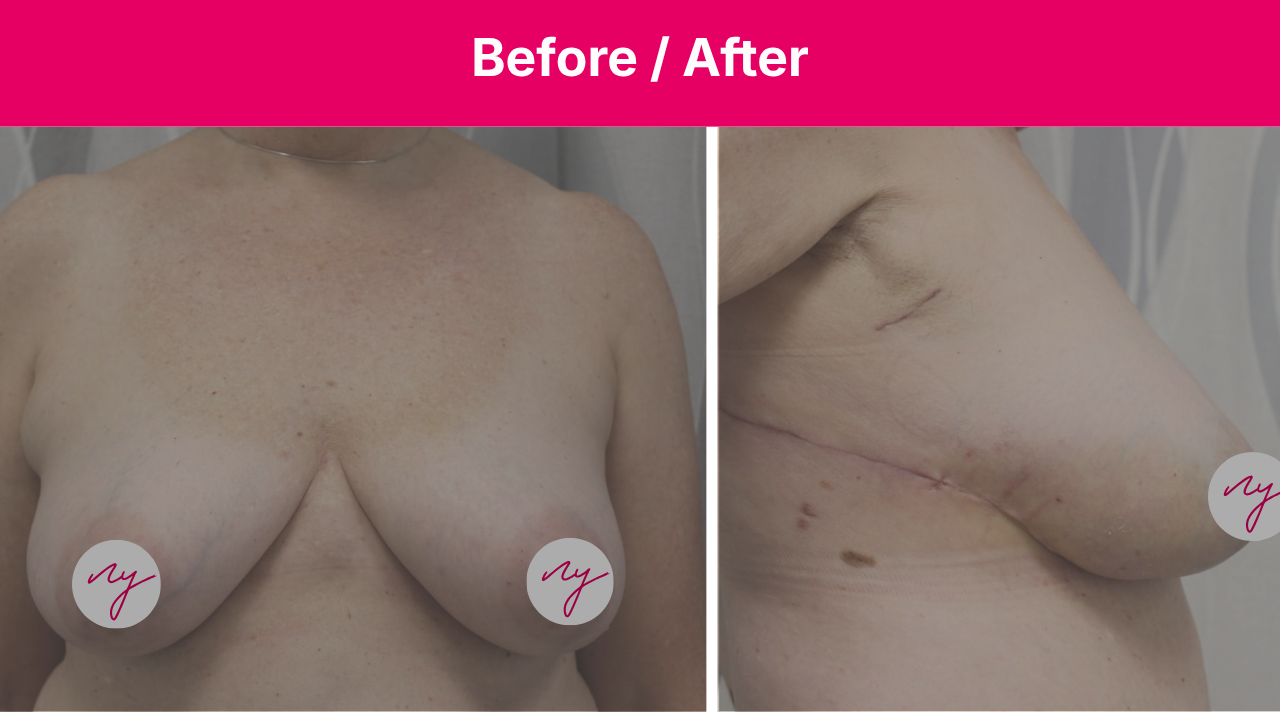
Preoperative photographs


Why a LICAP for this defect?
After discussing both options, the patient did not wish to undergo surgery on her other breast. As the tumour was located in an area particularly well suited to partial breast reconstruction with a chest wall perforator flap, she opted for this approach.
Tumours located in the lower outer quadrant of the breast are often particularly suitable for this type of reconstruction. The perforating vessels are located close to the defect created by tumour excision (see image below), allowing the flap to be rotated into the cavity through a short, tension-free arc. The donor site is positioned along the lateral chest wall, resulting in a well-hidden scar along the bra line.
Unlike a latissimus dorsi (LD) flap, a LICAP flap does not require sacrificing any back muscle. This preserves the musculature and avoids the potential functional morbidity associated with muscle harvest.
Key indication. The LICAP is best suited for defects in the lateral hemisphere of the breast where volume replacement (rather than displacement) is required, and where the tumor-to-breast-volume ratio would otherwise produce an unacceptable cosmetic result after simple wide local excision.

Post Operative Recovery
Recovery following a chest wall perforator flap is usually relatively quick, as no muscle is harvested or divided.
Patients are mobilized immediately after surgery and typically are discharged the next day with a postoperative drain for a few days. Any postoperative discomfort can usually be managed with standard pain medication.
Because the flap uses skin and fat while preserving the underlying muscle, recovery is generally less demanding than with muscle-based reconstruction.
Post Operative Outcome
Long-term patient satisfaction following LICAP reconstruction is generally high.
The donor-site scar can usually be concealed by a bra. Usually, there are no additional scars on the breast itself. Because the back muscles are preserved, there is also less concern about long-term shoulder or back functional impairment compared with muscle-based flaps.
For appropriately selected patients, LICAP can therefore provide durable breast reconstruction with good aesthetic and functional outcomes.


The LICAP is best suited for defects in the lateral part of the breast where volume replacement (rather than displacement) is required, and where the tumor-to-breast-volume ratio would otherwise produce an unacceptable cosmetic result after simple wide local excision.
Leave a Reply