
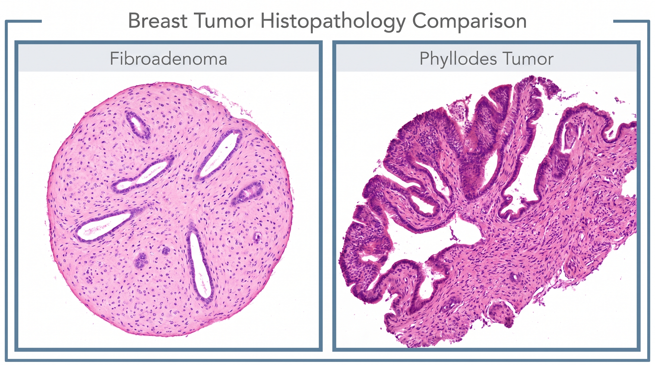
Two fibroepithelial lesions sit at the top of every breast clinic differential when a young woman presents with a palpable, well-defined lump. Here’s how to actually tell them apart — and why it matters.
breast-cancer breast-reduction breastcancer breastreduction cancer cancer-surgery chest wall perforator flap fibroadenoma health licap lumpectomy lymphnode surgery oncoplastic surgery partial breast reconstruction patient education phyllodes tumor sentinel node surgery surgery targeted axillary dissection therapeutic mammoplasty
When a young woman presents with a palpable, well-defined lump, the question arises. Is it a fibroadenoma (benign, common, reassuring) or a phyllodes tumor (rarer, occasionally malignant, occasionally terrifying)? The problem is, they look almost the same on ultrasound. Even under a microscope, they have similar appearances.
So how do you actually tell them apart — and why does it matter?
This post walks through the clinical, radiologic, histologic, and surgical differences that change management.
The Two Lesions at a Glance
Fibroadenoma
This is the most common benign breast lump. It usually feels like a smooth, rubbery bump that moves easily under the skin. It’s most often found in women under 35, though it can happen at any age. Many shrink or go away after menopause, and in most cases, they don’t need to be removed.
Phyllodes Tumor
A very rare tumor from the same fibroepithelial origin, but behaves differently. Under the microscope, it has a distinct leaf-like pattern (which is where the name comes from—phyllon means “leaf” in Greek). They can be classified as benign, borderline, or malignant. They can occur at any age but are most common in women between 40 and 50. The classic warning sign is a long-standing lump—often thought to be a fibroadenoma—that suddenly starts growing quickly.
A "fibroadenoma" in a 45-year-old that has doubled in size over three months should raise suspicion for phyllodes — even if prior biopsies said fibroadenoma.
Clinical Presentation
Fibroadenoma
- Painless, smooth, mobile, “breast mouse”
- Often discovered incidentally or on self-exam
- Slow-growing, may fluctuate with hormones
- Can be multiple in some patients
Phyllodestumor
- Larger at presentation (often 4–7 cm, sometimes >10 cm)
- Rapid growth over weeks to months
- Usually solitary
Imaging Findings
The initial investigation is usually an ultrasound and/or mammogram. In practice, both lesions are very similar on both mammography and ultrasound. Because imaging cannot reliably distinguish them, triple assessment (imaging + clinical history + tissue biopsy) is the gold standard.
Fibroadenoma

Phyllodes tumor

Images from https.radiopaegia.org
Breast biopsy — first step to give us potential answers
A core biopsy is the first step for a potential diagnosis. However, a tissue sample can underestimate a phyllodes tumor. It may sample a benign fibroepithelial area and miss the malignant stroma. Therefore, if a lesion is > 2-3 cm, growing rapidly, and the pathologist report cannot rule out phyllodestumor, a surgical excision is often recommended rather than observation, because the final pathology may upgrade to a phyllodes tumor.
Why do we need the tissue biopsy before removal?
When a biopsy confirms a small, asymptomatic fibroadenoma, conservative management with interval monitoring is the standard approach.
In contrast, any suspicion or histopathological confirmation of a phyllodes tumor mandates wide local excision with clear (negative) margins. The primary concern with phyllodes tumors is local recurrence, which is highly dependent on achieving adequate surgical margins.
Axillary staging is not routinely performed, as these tumors do not typically spread to regional lymph nodes.
Management Algorithm (Simplified)
Fibroepithelial lesion on biopsy │ ├─ Classic fibroadenoma features (young, <3 cm) │ → Observe or excision │ ├─ Cellular fibroepithelial lesion / features concerning │ → Diagnostic excision │ └─ Definite phyllodes → Wide local excision → Mastectomy if size/breast ratio demands it → No axillary dissection → Radiation considered for borderline/malignant with close margins → Chemo only for metastatic malignant disease
Key Takeaways
- Clinial presentation is your best guidance. Rapidly enlarging mass in a 40-year-old = differential diagnosis phyllodes.
- Core biopsy as first step.
- When there is doubt, excise.
- Margins matter. A wide local excision is needed for most phyllodes tumors.
- No axillary staging.
Leave a Reply